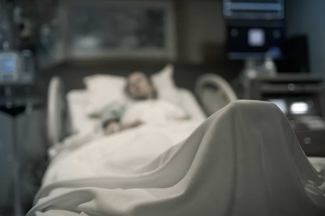
Failure to detect and respond to patient clinical deterioration is associated with increased hospital mortality and risk of adverse events that are known to be preventable.
Early recognition and response to deteriorating patients by health professionals is fundamental for optimal patient outcomes. On the contrary, delayed identification and management of deterioration can lead to serious consequences. Nurses play a pivotal role that requires appropriate and effective skills in this field and are required adequate and effective skills because they are the primary caregivers for deteriorating patients. This chapter aims to provide the essential skills for nurses and nursing students to confidently recognise and manage deteriorating patients in the acute setting.
Keywords: Detecting, Recognition, Response, Patient deterioration, Clinical Deterioration, Primary survey, ABCDE approach, Warning sings, Rapid Response.
-
Introduction
Early recognition and response by health professionals to patients whose physiological condition is acutely deteriorating is fundamental for safe and high-quality care, ensuring patients’ optimal outcomes. A timely, effective, and appropriate response may result in minimal intervention to stabilise the patient. The study demonstrates that prompt, efficient, and adequate response to patients reduces mortality, cardiac arrests, and hospital or ICU length of stay. Evidence suggests that there has been a growth in the application of the recognition and response systems for acute physiological deterioration.2 The Australian Commission for Quality and Safety in Health Care (ACQSHC) has stated the fundamental concepts of this approach as follows1:
- Recognition and response systems must always apply to all patients and should promote effective action by clinicians working in the wards and the accompanying medical officer or team. This includes requesting emergency assistance when necessary.
- Recognising and responding to acute physiological deterioration effectively necessitates clear communication of the diagnosis and overall treatment goals. This includes documenting information in the medical record and sharing information during clinical handovers and regular clinical rounds.
- Recognising and responding to acute physiological deterioration requires the availability of appropriately qualified, skilled, and experienced staff.
- Care should be tailored to the patient’s needs and expressed preferences, including previously documented advanced care plans and care objectives. If a patient lacks the capacity to engage in decision-making about their care, the views of a substitute decision-maker should be sought whenever possible.
The introduction of ACSQHC standard in Australia has resulted in an improvement in the outcome of hospitalised patients: More than 110 Australian Intensive Care Unit (ICU) equipped hospitals with the implication of the standard decreased cardiac arrest-related ICU admissions (from 5.6% to 4.1%) resulting in a 21% reduction in in-hospital mortality for cardiac-arrest related ICU admissions from the ward.3 Nurses, as the primary caregivers take part in and play a pivotal role in this field while caring for deteriorating patients. In this chapter, we discuss key elements regarding recognising clinical deterioration, which include Primary Survey (ABCDE approached assessment), Full Vital Signs, and Early Warning Signs.
-
Recognition of clinical deterioration
Research has consistently shown that there are observable and measurable physiological abnormalities prior to adverse events such as cardiac arrest, unanticipated admissions to intensive care and unexpected death.4-6
Early detection and intervention for clinical deterioration in a patient are critical, and they can be accomplished by implementing appropriate skills.
2.1 ABCDE assessment
The mnemonic “ABCDE” stands for airway, breathing, circulation, disability, and exposure. The ABCDE (Primary Survey) approach is a useful clinical tool for the initial assessment of rapidly deteriorating patients in an acute clinical setting. It has been designed to ensure that life-threatening conditions are identified and treated early and that there is an order of priority. If a problem is discovered as this clinical pathway is being implemented, the problem must be addressed immediately before moving on to the next part of the assessment.7,8
The structured ABCDE approach outlines each component that needs to be assessed briefly, which includes a patent’s airway and cervical spine stabilisation, breathing and ventilation, circulation with haemorrhage control, disability (neurological status), and exposure and temperature control.
The outcome of the assessment generates a general impression about the seriousness of the patients’ situation and how they are or may be affected.9,10 During the assessment, by looking, listening, and feeling, all the actual and imminent life-threatening risks, injuries, or illnesses, such as obstructed airways, fractures, brain injuries, haemorrhage, tamponade, and so forth, need to be identified, treated, or eliminated, and stabilised immediately to prevent further deterioration.11 At the end of the primary survey, the patient is completely exposed to a warm environment to ensure that there are no immediate-life-threatening injuries that have been missed for intervention, as well as to be prepared to proceed to a secondary survey. 9,11
A – Airway assessment
Is the airway patent? A primary survey begins by assessing and maintaining the patient’s airway, including checking airway patency, looking for obstructions and neck injuries, and, if indicated, providing cervical spine protection. Untreated, airway obstruction causes hypoxia and risks damage to the brain, kidneys, and heart, as well as cardiac arrest and death.11
This involves three steps:
- Look: If the patient can speak normally, move on to assessing the other ABCDEs. If the patient is unable to talk, look for airway obstruction (partial or full), symmetrical chest wall movement, or use of any accessory muscle such as abdominal movement.
- Listen and Feel: listen and feel for airflow at the mouth and nose.
B – Breathing
Is the breathing sufficiently maintained? This step is to assess the patient for any life-threatening respiratory conditions, exposing the chest and looking for signs of injury, measuring respiratory rate and oxygen saturation and assessing work of breathing and chest expansion.12,13
This involves three steps:
- Look: Observe symmetrical chest wall movement for any abnormalities, increased work of breathing, including the use of accessory muscles (neck and shoulders), nasal flaring, any signs of central cyanosis, and rate/depth of respiration.
- Listen: Does the patient speak in a short sentence? Auscultate the chest wall to detect any abnormal breath sounds such as wheeze, crackles, or stridor and to check for the absence of breath sounds.
- Feel: Feel the chest wall for surgical emphysema (possible indication of pneumothorax); the trachea alignment due to deviation suggests tension pneumothorax.
C – Circulation
Is the circulatory volume sufficient? It aims to control any external bleeding and identify any signs of hypovolemia from severe blood or fluid loss and shock by assessing heart rate and rhythm, blood pressure, and peripheral circulation.12,13
This involves three steps:
- Look: Look for signs of bleeding and poor circulation, such as pale or mottled skin.
- Listen (Pay attention): Does the patient appear confused? Does the patient have chest pain? These could indicate poor perfusion to the brain and heart due to decreased circulatory volume.
- Feel: Feel for signs of poor perfusions, such as cool, moist extremities, delayed capillary refill, diaphoresis, low blood pressure, tachypnoea, tachycardia, absent pulses, and low urine output.
D – Disability
What is the consciousness level? A quick evaluation of the patient’s conscious state and neurological function is performed as part of the evaluation of disability. Common causes of unconsciousness include profound hypoxia, hypercapnia, cerebral hypoperfusion, and the recent administration of sedatives or analgesics. During this phase, the patient’s blood glucose level should be checked to detect hypo- or hyperglycemia, which may contribute to altered mental status.13,14
Utilising the ACVPU scale (Table 1) will facilitate a rapid assessment of the patient’s level of consciousness, which will be followed by an assessment of the patient’s pupils (size, regularity, and responsiveness to light) and limb movement. Use the Glasgow Coma Scale if a more comprehensive assessment of the patient’s level of consciousness is required.
Table 1. The ACVPU scale measures a patient’s level of consciousness
| A | Patient is alert |
| C | New confusion or delirium |
| V | The patient is responding to verbal commands |
| P | The patient responds to a painful stimulus |
| U | The patient is unresponsive |
E – Exposure
Are there any other causes for the patient’s deterioration? The purpose of this is to investigate any clues that might explain the patient’s condition: It involves removing all of the patient’s clothing to inspect their body for any additional and potentially life-threatening injuries such as bleeding, rashes, bites, or other lesions, followed by promptly warming and maintaining control of the patient’s temperature. 12,13
2.2. Full Vital signs
To successfully recognise and respond to acute physiological decline, plans for monitoring vital signs and providing continuous patient care must be developed and communicated.1
Vital signs are a useful tool to identify patients who are at risk of deterioration because abnormal values in vital signs such as blood pressure (hypertension/hypotension), respiratory rate (tachypnoea/apnoea), heart rate (tachycardia/bradycardia), and low oxygen saturation often indicate patient deterioration prior to the occurrence of these serious adverse events [15]. To improve recognition of physiological abnormalities indicating deterioration, monitoring plans should at the very least include an assessment of the elements below and documentation in a structured tool, such as a paper or electronic observation and response chart.9
- Respiratory rate: Compare RR to previous measurements and search for deteriorating trends (Hyperventilation or Hypoventilation).
- Oxygen saturation: Compare the oxygen saturation level to previous measurements and search for deteriorating patterns (Hypoxia).
- Heart rate: Examine heart rate and compare with previous data, and search for deteriorating tendencies. If there are variations, how does the heart rate deviate from the usual heart rate? (Tachycardia or Bradycardia)
- Blood pressure: Compare blood pressure with previous values and search for deteriorating trends (Hypertension or Hypotension).
- Temperature: Check the temperature and compare it to previous measurements. Look for deteriorating trends (Hyperthermia or Hypothermia).
- Level of consciousness: Utilise A.V.P.U. (Alert, responds to Voice or Pain, Unconscious) to evaluate the patient’s mental status and any new-onset disorientation or behavioural change.
An increase in RR (Respiratory Rate) can usually be seen a few hours prior to a change in the other vital signs. As a result, even minor changes in RR can be a sign of a patient’s deterioration.16
Studies have shown that changes in a patient’s respiratory rate are reliable and predictive indicators of clinical deterioration in both adult and paediatric patients.17,18
As the body attempts to maintain homeostasis by delivering oxygen to organs and tissues, a deviation in respiratory rate can be the first sign of deterioration and an early indicator of physiological conditions that cause clinical deterioration such as hypoxia, hypercapnia, metabolic and respiratory acidosis. 19,20
2.3 Early warning sign
Early warning systems, commonly referred to as ‘track and trigger’ systems, are systematic processes that measure fundamental vital signs and respond to the results. When a predetermined criterion (the trigger) is met, an action should be taken. Many early warning systems rely on routine physiological monitoring, including vital signs. The calling criteria for a medical emergency team (MET) are one of the most common types of single parameter systems in Australia: With periodic monitoring of selected vital signs against a simple set of predefined criteria, with a response algorithm activated when any criterion is met.21
Table 2 lists the early and late signs reported as predictors of serious adverse events such as death, cardiac arrest, severe respiratory problems, and transfer to critical care in the Signs of Critical Conditions and Emergency Responses (SOCCER) studies.22 Early warning scores and Rapid Response Teams will be further discussed in response to the clinical deterioration section.
Table 2. Key criteria requiring further assistance with patient assessment and management.
| Criterions | Early warning signs of patient deterioration | Late warning signs of patient deterioration |
| Airway | Partial airway obstruction (excluding snoring) | Airway obstruction or stridor |
| Oxygen Saturation | SaO2 90–95% | SaO2 < 90% |
| Respiratory Rate | Respiratory rate 5–9 or 30–40/min | Respiratory rate < 5 bpm or > 40/min |
| Heart Rate | Pulse rate 40–50 bpm or 120–140bpm | Pulse rate < 40 bpm or >140bpm |
| Blood Pressure | Systolic BP 80–100 mmHg or 180–240 mmHg | Systolic BP < 80 mmHg or > 240 mmHg |
| Urine Output | Urine output < 200 mL over eight hours | Urine output < 200 mL in 24 hours or anuria |
| Drainage Output | Greater than expected drainage fluid loss | Excess blood loss not controlled by ward staff |
| Mental Status | A drop in GCS of 2 points or GCS < 12 or any seizure | Unresponsive to verbal commands or
GCS < 8 |
| ABGs | PaO2 50–60,
PCO2 50–60, PH 7.2–7.3 | PaO2 < 50,
PCO2 > 60, PH < 7.2, |
References
1 Australian Commission on Safety and Quality in Health Care (ACSQHC), 2021. Essential elements for recognising and responding to acute physiological deterioration (3rd Ed.). The NSQHS Standards. www.safetyandquality.gov.au/standards/nsqhs-standards
2 Bucknall T., McKinley, S., Fossum, M., & Austin, N. (2016).Rapid review of the literature and draft revision of the National consensus statement: essential elements for recognising and responding to clinical deterioration.
3 Martin C, Jones D, Wolfe R. State-wide reduction in in-hospital cardiac complications in association with introduction of a national standard for recognising deteriorating patient. Resuscitation. 2017; 121:172-178
4 Buist M, Bernard S, Nguyen TV, Moore G, Anderson J. 2004.Association between clinical abnormal observations and subsequent in-hospital mortality: a prospective study. Resuscitation. 62:137-41. 5.
5 Fuhrmann L, Lippert A, Perner A, Østergard D. 2008. Incidence, staff awareness and mortality of patients at risk on general wards. Resuscitation. 77(3):325-30.
6 Kause J, Smith G, Prytherch D, Parr M, Flabouris A, Hillman K, et al.2004. A comparison of Antecedents to Cardiac Arrests, Deaths and Emergency Intensive care Admissions in Australia and New Zealand and the United Kingdom – the ACADEMIA study. Resuscitation. 62:275-82.
7 Smith, D. (2020). Do you use your ABCDE? Nursing Standard, 35(3), 67-68. doi:http://dx.doi.org.ez.library.latrobe.edu.au/10.7748/ns.35.3.67.s23
8 Hill, R., Hall, H., & Glew, P. (2017). Fundamentals of Nursing and Midwifery A person-centred approach to care (3rd ed.) Wolters Kluwer Health.
9 Bowden, T. & Smith, D. (2017). Using the ABCDE approach to assess the deteriorating patient. Nursing Standard, 32(14), 51-61. doi:10.7748/ns.2017.e11030
10 Cameron, P., Little, M., Jelinek, G., Kelly, A. and Brown, A. (2014). Trauma. In Textbook of Adult Emergency Medicine Expert Consult-Online and Print (pp. 73-75). Saint Louis, UK: Elsevier Health Science. Retrieved from https://ebookcentral-proquest com.ez.library.latrobe.edu.au/lib/latrobe/reader.action?docID=1746435&ppg=98
11 Trauma Victoria. (2021). Retrieved from Primary Survey: https://trauma.reach.vic.gov.au/guidelines/early-trauma-care/primary-survey
12 Curtis, K., Ramsden, C., & Lord, B. (2011). Emergency and Trauma Care for Nurses and Paramedics. Elsevier Health Sciences
13 Victorian State Trauma System. (n.d.). Early Trauma Care. Trauma Victoria. Retrieved April 20, 2021, from https://trauma.reach.vic.gov.au/guidelines/early-trauma-care/primary-survey
14 Cameron, P., Jelinek, G., Kelly, A., Brown, A., & Little, M. (Eds.). (2015). Textbook of Adult Emergency Medicine (4th ed.). Elsevier Health Sciences.
15 Brekke, I. J., Puntervoll, L. H., Pedersen,P. B., Kellett,J., & Brabrand, M. (2019). The value of vital sign trends in predicting and monitoring clinical deterioration
A systematic review. Plos One, 14(1). doi: 10.1371/journal.pone.0210875
16 Rolfe, S. (2019). The importance of respiratory rate monitoring. British Journal of Nursing, 28(8), 504-508. https://doi.org/10.12968/bjon.2019.28.8.504
17 Daw, W., Kaur, R., Delaney, M., & Elphick, H. (2020). Respiratory rate is an early predictor of clinical deterioration in children. Pediatric Pulmonology, 55(8), 2041-2049. https://doi.org/10.1002/ppul.24853
18 Mochizuki, K., Shintani, R., Mori, K., Sato, T., Sakaguchi, O., Takeshige, K., Nitta, K., & Imamura, H. (2017). Importance of respiratory rate for the prediction of clinical deterioration after emergency department discharge: a single centre, case-control study. Acute Medicine & Surgery, 4(2), 172-178. https://doi.org/10.1002/ams2.252
19 Waugh, A., & Grant, A. (2014) Ross and Wilson Anatomy & Physiology (12th ed.). Elsevier: NSW
20 Flenady, T., Dwyer, T., & Applegarth, J. (2017). Accurate respiratory rates count: So should you. Australasian Emergency Nursing Journal, 20(1), 45–47. https://doi.org/10.1016/j.aenj.2016.12.003
21 Chatterjee, M. T., Moon, J. C., Murphy, R., & McCrea, D.(2005). The “OBS” chart: an evidence based approach to re-design of the patient observation chart in a district general hospital setting. Postgraduate Medical Journal. 81:663-6
22 Jacques, T., Harrison, G. A., McLaws, M-L., & Kilborn, G. (2006). Signs of critical conditions and emergency responses (SOCCER): A model for predicting adverse events in the inpatient setting. Resuscitation, 69(2):175-83.
Conflict of Interest
All the authors declare they have no conflict of interest in relation to this manuscript.
Authors – contributed equally to authorship
Seung A (Sarah) Park, is a Nursing Lecturer at Chisholm Higher Education, Critical Care Registered Nurse at St John of God hospital ICU.Taryn Kellerman, is a Nursing Lecturer at Chisholm Higher Education, District Registered Nurse at Bolton Clarke.


